Cards In This Set
| Front | Back |

|
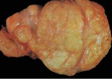
Fibroadenoma. Gross image.
The cut surface of this mass is characteristic for fibroadenoma with its slightly lobulated pattern and solid firm, bulging grayish-tan parenchyma. Occasional slit-like spaces can be seen. Myxoid change characterized by a glistening surface and calcification in longer standing lesions is not uncommon. Digital Atlas of Breast Pathology by Meenakshi Singh, MD © - Department of Pathology, Stony Brook University Medical Center/breast-atlas/XXVII-21.htm /topic/breastfibroadenoma.html |
 |
Fibroadenoma
Gross description (Macroscopy) ========================================================================= ● Sharply circumscribed with smooth, rounded border, freely movable spherical nodule, usually 3 cm or less ● Gray-white, bulging cut surface with numerous slits ● 20% multifocal http://health-pictures.co/images/Fibroadenoma.jpg/topic/breastfibroadenoma.html |
 |
Fibroadenoma
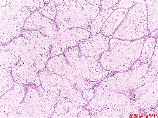
@font-face { font-family: "Arial"; }@font-face { font-family: "Times"; }@font-face { font-family: "Cambria"; }p.MsoNormal, li.MsoNormal, div.MsoNormal { margin: 0in 0in 0.0001pt; font-size: 12pt; font-family: "Times New Roman"; }div.Section1 { page: Section1; } @font-face { font-family: "Arial"; }@font-face { font-family: "Times"; }@font-face { font-family: "Cambria"; }p.MsoNormal, li.MsoNormal, div.MsoNormal { margin: 0in 0in 0.0001pt; font-size: 12pt; font-family: "Times New Roman"; }div.Section1 { page: Section1; } Micro description (Histopathology) ========================================================== ● Rounded contour, overgrowth of fibrous and glandular tissue ● Intralobular stroma (delicate, cellular, myxoid or fibrotic) encloses glandular spaces; may infarct, become inflamed, calcify ● Pericanalicular: open glandular spaces vs intracanalicular: compressed glandular spaces [no clinical significance to this distinction] ● Glands have cuboidal/low columnar epithelium and adjacent myoepithelium, but no atypia ● 15% have apocrine metaplasia ● May have myxoid change (suggests Carney’s syndrome), sclerosing adenosis, epithelial hyperplasia or other fibrocystic change ● May have adjacent fibroadenomatous change ● Rarely has pleomorphic, bizarre multinucleated giant cells (Arch Pathol Lab Med 2000;124:1721, Diagn Pathol 2008;3:33, Am J Surg Pathol 1986;10:823, Arch Pathol Lab Med 1994;118:912), squamous metaplasia, smooth muscle or adipose tissue, metaplastic cartilage, DCIS or LCIS ● Fibroadenoma phyllodes: rarely has focal leaf-like processes but otherwise typical fibroadenomatous stroma ● No necrosis, no elastic tissue, no anaplasia, no mitotic figures http://pathology.class.kmu.edu.tw/ch10/84-2.jpg/topic/breastfibroadenoma.html |

|
Fibroadenoma
Fibroadenoma with atypical lobular hyperplasia. Note that the lobules in the left side of the photomicrograph are filled with neoplastic cells, but there is no distention of the lobular units. Digital Atlas of Breast Pathology by Meenakshi Singh, MD © - Department of Pathology, Stony Brook University Medical Center /breast-atlas/XVII-03.htm /topic/breastfibroadenoma.html |

|
Fibroadenoma : Pericanalicular
Comments: The tubules and glands in a fibroadenoma are lined by cuboidal or low columnar epithelium with uniform nuclei and surrounded by a myoepithelial layer. The stroma is made up of loose connective tissue. If the stroma is hypercellular, the diagnosis of phyllodes tumor should be excluded. http://webpathology.com/image.asp?case=276&n=7 /topic/breastfibroadenoma.html |

|
Intraductal Papilloma of Breast
Comments: The image shows an intraductal papilloma arising in a small duct. Such lesions are microscopic and not detected on palpation. The average age at presentation is 48 yrs. /image.asp?case=280&n=1 |

|
Intraductal Papilloma of Breast
Comments: Architecturally, papillomas show complex, intricate branching fronds that are lined by a layer of epithelium consisting of cuboidal or columnar cells and myoepithelial cells. http://webpathology.com/image.asp?case=280&n=5 |
 |
Intraductal papilloma.
Papillary proliferation with irregular glandular spaces.
|

|
Intraductal Papilloma of Breast
Comments: This image shows a 1.5 cm intraductal papilloma that created a palpable subareolar mass accompanied by bloody nipple discharge. Most larger intraductal papillomas are solitary. http://webpathology.com/image.asp?case=280&n=3 |

|
Central papilloma with florid, ductal hyperplasia of the usual type.
Digital Atlas of Breast Pathology by Meenakshi Singh, MD © - Department of Pathology, Stony Brook University Medical Center/breast-atlas/XIII-06B.htm |

|
Sclerosing Adenosis of Breast
Comments: Sclerosing adenosis is a benign hyperplastic process that may be mistaken for carcinoma. The average age at presentation is about 30 yrs. The lesion retains is lobular configuration and is more cellular centrally. http://webpathology.com/image.asp?n=1&Case=281 |

|
Sclerosing Adenosis of Breast
Comments: The proliferating tubules may be elongated and have attenuated lumens. There is preferential preservation of myoepithelial cells in the tubules and epithelial cells are less conspicuous. Some degree of lobular fibrosis is usually present. http://webpathology.com/image.asp?n=2&Case=281 |

|
Sclerosing Adenosis of Breast
Comments: Myoepithelial component (cells with clear cytoplasm in this image) may predominate in some cases of sclerosing adenosis. Their presence can be confirmed with immunohistochemical stains (smooth muscle actin, calponin, p63). http://webpathology.com/image.asp?n=3&Case=281 |

|
Sclerosing Adenosis of Breast
Comments: Some cases of sclerosing adenosis don't have lobulocentric architecture and may have infiltrative edges. This may lead to the mistaken diagnosis of well-differentiated ductal carcinoma, especially in limited needle core biopsy specimens. http://webpathology.com/image.asp?n=4&Case=281 |

|
Sclerosing adenosis
A low power view with the proliferative process retained within essentially lobular configurations is important. If questions arise about the invasive nature high molecular weight cytokeratin staining of myoepithelial cells (i.e. P63) can be invaluable. Digital Atlas of Breast Pathology by Meenakshi Singh, MD © - Department of Pathology, Stony Brook University Medical Center /breast-atlas/V-27.htm |


