Cards In This Set
| Front | Back |
|
Fatty Infiltration
|
Benign, reversible disorder that occurs when fat is deposited in liver, mostly right lobeLiver typically enlarged and echogenicTypical fat-spared area anterior to right portal vein
|
|
Cirrhosis
|
Irreversible disease that leads to loss of normal liver function and death of hepatocytesIn long term, liver failure and portal hyptertensionEarly stages, liver enlarged by hepatitisLater stages, fatty infiltrationFinal stage, nodular normal/small liver often outlined by ascites; portal hypertension
|
|
Portal hypertension
|
Occurs when intrahepatic processes lead to increased port vein pressureNormal flow hepatopetalReversed hepatofugalCollaterals develop, i.e. recanalized paraumbilical veins (umbilical v. in ligamentum teres)SplenomegalyIn porta hepatis, if pv is thrombosed, collaterals form as cavernous transformation
|
|
Hepatitis
|
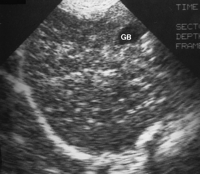
 Self-limiting: acute viral3-6 months: chronicLeads to cirrhosis and liver failureApparent increase in portal vein echogenicity caused by decrease in liver echogenicity ("starry night" appearance)Hepatosplenomegaly |
|
Budd-Chiari syndrome
|
Occlusion of hepatic veins w/ or w/out occlusion of IVCAscites always seenHepatomegalyCD of hepatic veins will show absence of veins, intravenous thrombi, intrahepatic collaterals, variable vein size
|
|
Cavernous Hemangioma
|
Most common benign liver tumorVascular lesions seen more frequently in womenUsually in right lobeSmall echogenic lesion that may display post enhancementUsually well-defined borders with slightly less echogenic center
|
|
Adenomas
|
Benign, seen more frequently in womenLinked to use of oral contraceptivesCan be hypoechoic or hyperechoic
|
|
Focal Nodular Hyperplasia (FNH)
|
Benign tumor, more common in womenMay be hypoechoic or hyperechoicWell-defined border and central scar
|
|
Hepatocellular Carcinoma (Hepatoma)
|
More common in menUsually associated with macronodular liver cirrhosis and alcoholismSolitary or mult nodules or diffuse infiltrationEchogenic or complexMay invade portal vein and mimic portal vein thrombosis
|
|
Hepatoblastoma
|
Rare malignant tumorMost common liver mass in infantsRaised alpha-fetoprotein levelsHighly vascular solitary massPossible calcifications
|
|
Metastases
|
Very common in liverPrimary sites:Lymphoma, breast, lung: hypoechoic massesKidneys, pancreas: echogenic massesColon, pancreas, stomach, neuroblastoma: calcified massesColon, lung: masses with a "bull's eye" or "target" appearance
|
|
Cysts
|
Acquired or congenitalMaybe be single or mult, and unusual shapes are commonSame features as elsewhere in the body aside from shape
|
|
Hydatid (Echinoccocal) Cysts
|
Usually related to tapeworm in dogs; seen more frequently in sheep- and cattle-raising areasIncreased WBC countUsually in right lobeCyst within a cyst often seen ("Water Lily")May have mass-like appearance with internal echoes or may become calcified as it ages
|
|
Pyogenic Abscesses
|
May be secondary to diverticulitis, appendicitis, cholecystitis, or surgeryAerobic and anaerobic organisms found in itComplex fluid collection with good through transmissionIrregular, thick wall
|
|
Amebic Abscesses
|
Most frequently in tropical regionsCaused by protozoa Entamoeba, or poor sanitary conditionsRound, hypoechoic or hyperechoic mass
|


