Related Flashcards
Related Topics
Cards In This Set
| Front | Back |
|
The maximum useful magnification for a light microscope is...
|
1000x
|
|
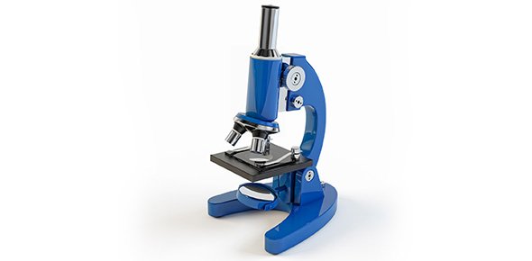
The 2 image forming lenses of a compound light microscope are...
|
Ocular and objective
|
|
Dyes are usually added to sections of biological specimens to increase...
|
Contrast
|
|
What kind of microscope do you use if observing living surface of a finger?
|
Scanning Electron Microscope (SEM)
|
|
What kind of microscope is used for observing a dye stained slide of a section of your finger?
|
Compound Light Microscope
|
|
What microscope is used for observing gold coated bacteria on a single cell of a finger?
|
Scanning Electron Microscope (SEM)
|
|
What microscope is used when observing a unstained section of a biopsy from a finger?
|
Compound Light Microscope
|
|
WHat microscope is used when observing a heavy-metal-stained very thin section of a finger?
|
Transmition Electron Microscope (TEM)
|
|
Forms a TV Like picture from a secondary electron signal, which is emitted from the surface points excited by a thin beam of electrons drawn across the surface in a raster pattern.
Investigates fine structure of surface. (Usually dead specimens)
|
Scanning Electron Microscope
|
|
Increases resolving power by using electrons in a vacuum and magnetic lenses instead of light and glass lenses.
Preservation of greater specimen detail allows for magnifications up to 1,000,000x or more (usually dead materials)
|
Transmission Electron Microscope
|
|
Converts phase differences in light to differences in contrast
Observation of low contrast specimens (often Living)
|
Phase-Contrast and Nomarski Process
|
|
Four steps when using compound light microscope.
|
1. Carry it to and from lab table by grasping arm and supporting base.
2. Keep it upright
3. Remove dust cover
4. If it needs to be dusted, use lens paper only.
|
|
The amt that an object image is enlarged
|
Magnification
|
|
The extent to which detail in an image is preserved during the magnifying process
|
Resolving Power
|
|
Focusesradiation emanating from an objective to produce an image
|
LEns
|


