Related Flashcards
Related Topics
Cards In This Set
| Front | Back |
|
Chapter 9: The Development of the Ectoderm
How the Neural tube and Epidermis Arise and Acquire their Distinctive Patterns Ectoderm is one of the three primary germ cell layers in the very early embryo; it is the most exterior (distal) layer and differentiates into the nervous system. What are the 3 major derivatives of the ectoderm germ layer? Which derivative of the ectoderm germ layer is the foundation of neurluation? Major Derivatives of the Ectoderm Germ Layer Epidermis, Hair, Nails, olfactory epithelium, mouth epithelium, sebaceous glands, lens and cornea are part of which major derivative of the ectoderm germ layer? Periphery nervous system, adrenal medulla, melanocytes, facial cartilage and dentine of teeth are part of which major derivative of the ectoderm layer? Brain, spinal cord, retina, motor neurons, neural pathway |
Surface ectoderm, neural crest and neural tube
Surface Ectoderm Neural Crest Neural Tube |
|
Chapter 9: The Development of the Ectoderm
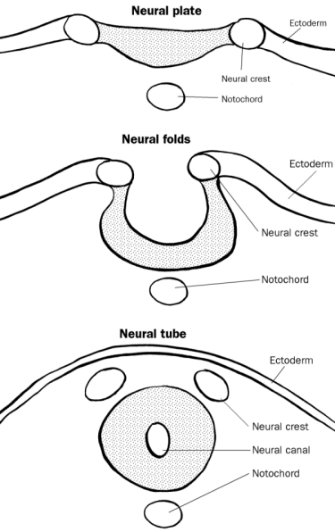
How the Neural tube and Epidermis Arise and Acquire their Distinctive Patterns Primary Neuralation~ Neural Tube Fomation in Chick Embryo Primary neurulation is the conversion of the ______ into the _____. Folding begins as the _____ (MHP) cells anchor to the notochord and change their ____, while the presumptive epidermal cells move toward the dorsal midline. Next, the neural folds are _____ while the presumptive epidermal folds continue to move toward the dorsal midline. The neural folds are brought into contact with one another----what links the neural tube wih the epidermis? What separates the neural tube from the epidermis? |
Neural plate; neural tube
Medial Hinge point; shape elevated neural crest Dispersion of neural crest |
|
Chapter 9: The Development of the Ectoderm
How the Neural tube and Epidermis Arise and Acquire their Distinctive Patterns Neurulation in Human Embryo The anterior neural pore is usually closed by day 25 or 26; the failure to close this pore leads to what condition? Anencephaly is considered what kind of defect? What are the plausible causes? Expression of N and E-Cahedrin Adhesion Proteins What are the normal expressions of N-Cahedrin and E-cahedrin? What happens when N-Cahdrin is over-expressed? Folate Binding Protein Folate appears to be critical in human neural closure---what have recent studies demonstrated expression of immediately prior to fusion? |
Anencephaly
Neural tube defect Environmental toxins; low intake of folic acid during pregnancy N-Cahedrin is normally expressed in the neural plate and E-Cahedrin is normally expressed in the epidermis If N-Cahedrin is over-expressed neural tube separation does not occur folate receptor protein |
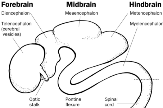
 Chapter 9: The Development of the Ectoderm How the Neural tube and Epidermis Arise and Acquire their Distinctive Patterns Secondary Neurulation What do mesenchymal cells have to do to form the medullaly cord beneath the surface ectoderm? What type of transition is this? After this transition what does the central portion of the medullary cord undergo and what does it form? How is the central canal of the neural tube formed? Early Human Brain Development Differentiation of the neural tube into the various regions of the ______ nervous system occurs simultaneously in 3 different ways. Name the 3 cavities/ primary brain vesicles. When do these cavities form? What does the prosencephalon (forebrain) subdivide into? What does the midbrain (mesencephalon) subdivide into? What does the Rhombencephalon subdivide into? Which of the 5 secondary vesicles forms the cerebral hemisphere? Which forms the optic vesicles (future retina) as well as the thalmamic and hypothalamic brain regions? Which subdivision of the hind brain becomes the medulla oblongota? Which gives rise to the cerebellum? |
Condense
Mesenchymal to Epithelial Cavitation; forms lumen (hollow spaces) The lumen coalesce into a single cavity central Forebrain (Prosencephalon) Midbrain (Mesencephalon) Hindbrain (Rhombencephalon) During embryonic development of the brain Telencephalon and Diencephalon Does not subdivide, remains mesencephalon Metencephalon and mylencephalon Telencephalon Diencephalon Mylencephalon Metencephalon |
|
Chapter 9: The Development of the Ectoderm
How the Neural tube and Epidermis Arise and Acquire their Distinctive Patterns Dorsal And Ventral Specification of Neural Tube There are 2 signals that influence the neural tube: What is the roof of the neural tube exposed to? What exposes the neural tube to BMP4 and BMP7? What is the floor plate exposed to? What exposes the floor to this protein? Both the TGF-beta family (BMP4 & 7) and SHH form gradients in the neural tube; which spreads ventrally (back to belly---ventral end)? which spreads dorsally (Belly to back---dorsal end)? Exposure to the gradients of paracrine factors gives which neurons their identites? The gradient of Sonic hedge- hog specifies the ventral neural tube by activating and inhibiting the synthesis of particular transcription factors. |
BMP4 & BMP7
The epidermis Sonic Hedge Hog Notochord TGF-Beta spreads ventrally (back to belly) SHH spreads dorsally (belly to back) spinal cord |
 |
Embryonic Brain structure
|
 |
Neural plate----Neural Folds-----Neural Tube
|
|
Neural Crest cells are pleuripotent and migrate widely throughout the embryo giving rise to many nervous structures.
|
Overview
|
|
Chapter 9: The Development of the Ectoderm
How the Neural tube and Epidermis Arise and Acquire their Distinctive Patterns Neural Stem Cells In the neural tube, where are neural stem cells in the mitotic phase found? Review of the Cell Cycle In G1, what happens to the celluar contents (except the chromosomes)? In the S phase what is duplicated/replicated? What is the purpose of the second gap phase? |
Near the lumen
Cellular contents are duplicated Genome is replicated Check for errors and make repairs |
|
Chapter 9: The Development of the Ectoderm
How the Neural tube and Epidermis Arise and Acquire their Distinctive Patterns Differentiation of the walls of the Neural Tube The neural tube consists of three cellular layers from inner to outer: Name them. What types of cells does the ventricular zone give rise to? Where do these cells migrate? In the spinal cord and medulla, which zone remains the sole source of neurons and glial cells? |
Ventricular zone, intermediate zone and marginal zone
Neuroblasts and gioblasts Migrate to the intermediate zone Ventricular zone |
|
Chapter 9: The Development of the Ectoderm
How the Neural tube and Epidermis Arise and Acquire their Distinctive Patterns Differentiation of the walls of the Neural Tube: Cerebrellar Organization In the cerebellum some neuroblasts migrate to the region farthest removed from the ventricular zone. They form a second mitotic layer----what is it called? The neuroblasts come in contact with BMP factors, what do they specify the neuroblasts into? These granule cells migrate back to the ventricular zone to form what layer? Meanwhile, the original ventricular zone of the cerebellum generates a wide variety of neurons and glial cells, including the distinctive and large Purkinje neurons, the major cell type of the cerebellum. What protein do these neurons secrete?What does Sonic Hedge Hog sustain? SHH is secreted by the notochord and participates in _____ signaling. BDNF is also a paracrine factor and regulates the _____ of migration. Which layer is made by? BMP is a member of the TGF beta family and specifies neuroblasts into ____ cells. |
External granular layer
Granule cells internal granular layer Sonic Hedge Hog Sustains the division of granule cell precursors Paracrine direction internal granular layer granule |
|
Chapter 9: The Development of the Ectoderm
How the Neural tube and Epidermis Arise and Acquire their Distinctive Patterns Differentiation of the walls of the Neural Tube: Cerebral Cortex The migrating neuroblasts and glioblasts generated in the ventricular zone migrate outward along radial glial processes to form what? How many layers does this plate have? The fates of neuronal precursors from OLDER brains are more ____. While the neuronal precursor cells formed EARLY in development have the potential to become any neuron (at layers 2 or 6, for instance); later precursor cells give rise only to upper-level (layer 2) neurons. Which neurons migrate faster to further layers? A neuron's birthday is the day that a neuron divides into a ____ cell (residing in the ventricular zone). |
Cortical plate
6 fixed older stem |
|
Chapter 9: The Development of the Ectoderm
How the Neural tube and Epidermis Arise and Acquire their Distinctive Patterns Differentiation of the walls of the Neural Tube: Cerebral Cortex Corticlal neurons are generated from 3 types of precursor cells: name them. Which precursor cell do RGCs give rise to? Which precursor cell(s) divides at the luminar surface of the ventricular layer? Which divide away from the luminar surface? Which zone (ventricular or subventricular) generates lower layer neurons? Upper layer neurons? |
RGC, SNPs and IPCs (Radial Glial Cells, Short neural precursors and intermediate precursor cells)
IPCs RGCs and SNPs IPCs ventricular; subventricular |
|
Chapter 9: The Development of the Ectoderm
How the Neural tube and Epidermis Arise and Acquire their Distinctive Patterns Both human and ape brains have a high growth rate before birth. After birth, however, this rate slows greatly in the ____, whereas human brain growth continues at a rapid rate for about 2 years. |
Apes
|
|
Chapter 9: The Development of the Ectoderm
How the Neural tube and Epidermis Arise and Acquire their Distinctive Patterns Development of the Vertabrate Eye The vertabrate eye is formed by interaction of the neural tube with a series of epidermal thickenings----what is this called? Which 3 transcription factors are involved in the development of the optic vesicle? Which plays a role in the specification of the lens and retina? Noggin inhibits ____ expression but promotes expression of _____. Otx2 protein then represses the inhibition of ET by Noggin signaling. The ET transcription factor activates _____, which encodes a transcription factor that blocks Otx2 and promotes ____ expression. What does pax6 initiate? Which protein (not mentioned above) is responsible for separation of the single eye field into 2 bilateral fields? What happens if SHH is mutated or processing is inhibited? |
Cranial ectoderm placodes
Six3, Pax6, Rx1 ET; OTx2 Rx1; Pax6 Initiates a gene cascade contributing to eye field SHH The single eye field does not split and results in cyclopia |


