Anatomy And Physiology Lab 2

Total Flash Cards » 35
Text Size: S | M | L

Bony Joints

synostosis
synarthrotic
ossification of fibrous or cartilaginous joints
Fibrous Joints Synarthrotic
or synarthrodial joint
adjacent bones are bound by collagen fibers that emerge from one bone
sutures, gomphoses, and syndesmoses
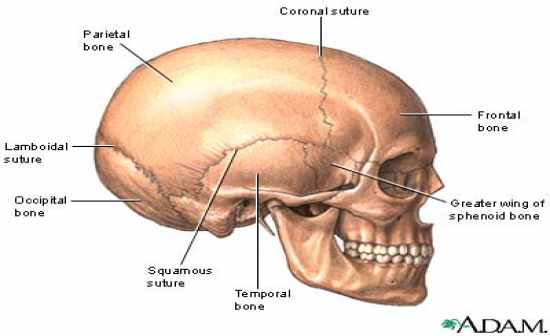
sutures synarthrotic

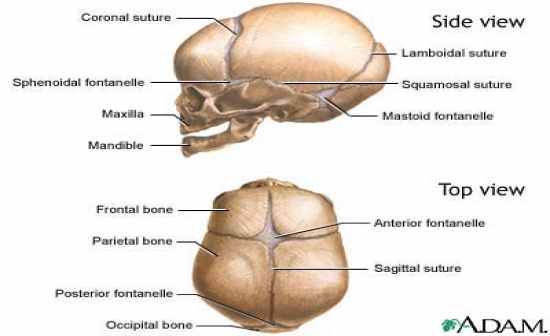
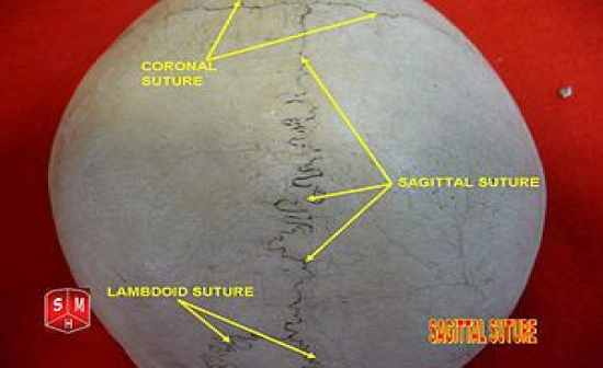
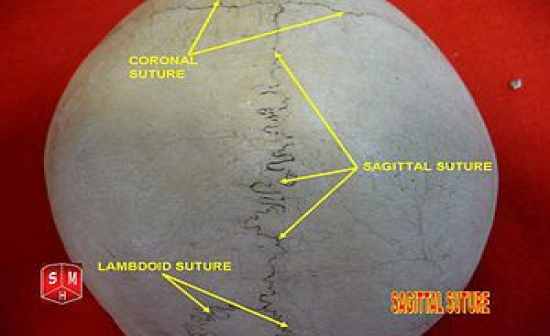
sagittal suture
between parietal bones

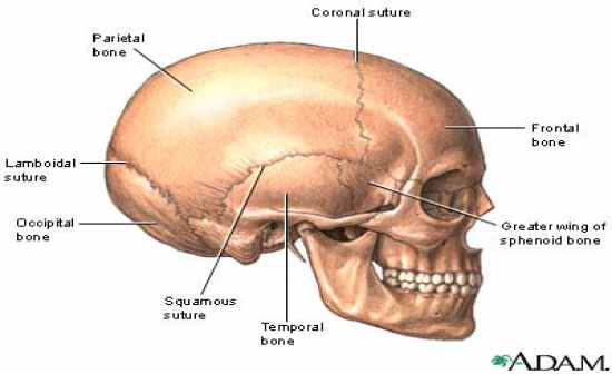
coronal suture
at the anterior margin

lambdoid suture
at the posterior margin

squamous suture
laterally

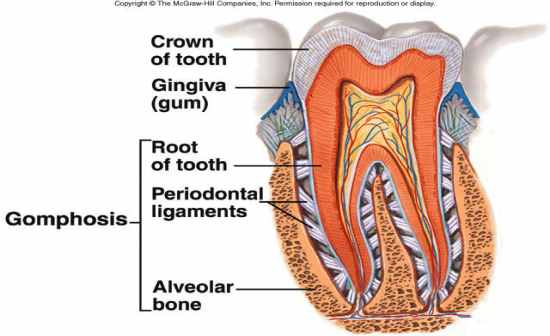
gomphoses
synarthrotic

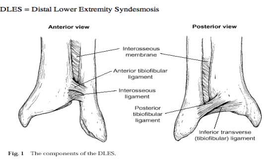
syndesmoses
amphiarthrotic
Cartilaginous joints amphiarthrosis
two bones linked by cartilage

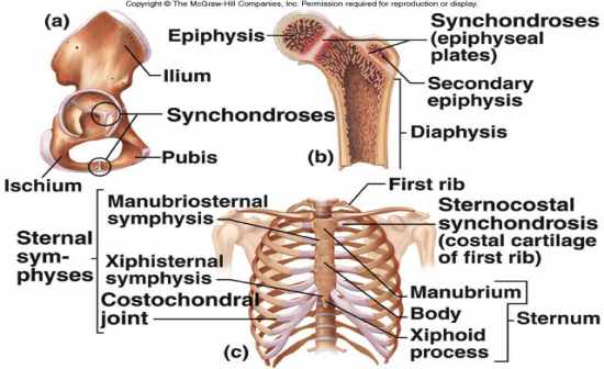
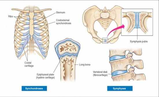
Synchrondroses
synarthrotic
bones are bound by hyaline cartilage
between epiphysis and diaphysis of a long bone
first rib to the sternum by a hyaline costal cartilage

symphyses
amphiarthrotic
two bones joined by fibrocartilage
pubic symphysis- R and L pubic bones are joined by the cartilaginous interpubic disc
synovial joints diarthrotic

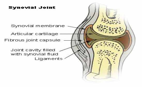
articular cartilage
in synovial joints
facing smooth surfaces of the two bones are covered in this
hyaline cartilage

joint (articular cavity)
synovial fluid
joint (articular) capsule
fibrous capsule
synovial membrane
--

spheroid joints
shoulder and hip joints
multiaxial joints
the glenoid cavity of the scapula
acetabulum of the hip bone
ellipsoid joints oval convex surface on one bone that fits into a complementary shaped depression on the other
radiocarpal joint of the wrist and metacarpophalangeal joints at the bases of the fingers
biaxial
sellar joints saddle-shaped surface-concave in 1 direction and convex in the other
trapeziometacarpal joint between trapezium of the wrist and metacarpal 1 at the base of the thumb
biaxial

plane (gliding) joints
arthodial
flat articular surfaces where bones slide over each other
limited movement
carpal bones of the wrist
tarsal bones of ankle
articular processes of the vertebrae
biaxial

ginglymus joints
monaxial joints
1 bone with convex surface that fits into a concave depression on the other bone
elbow, knee, and interphalangeal (finger and toe) joints

trochoid joints
monaxial joints
spins on a longitudinal axis
atlantoaxial joint between the 1st 2 vertebrae
radioulnar joint at elbow

flexion and extension
flexion: decreases a joint angle (usually sagittal plane)-hinge joints
extension: movement that straightens a joint and generally returns a body part to the zero position- straightening elbow

abduction and adduction
abduction: is a movement of a body part in the frontal plane away from the body
adduction: movement in frontal plane back toward the midline

Elevation and Depression
elevation: movement that raises a body part vetically in the frontal plane
depression: lowers a body part in the same direction

protraction and retraction
protraction: anterior movement of a bady part in the transverse (horizontal) plane
retraction: posterior movement

Circumduction
one end of an appendage remains fairly stationary while the other makes a circular motion

rotation
bone turning around an axis
--> movement in which a bone spins on its longitudinal axis
medial rotation: humerus spins
lateral rotation: opposite motion, so forearm points away from the body

pronation and supination
supination: of the foremarm is a movement that turns the palm to face anteriorly or upward SAP: forearm is supinated and the radius is parallel to the ulna
Pronation: is the opposite movement causing the palm to face posteriorly or downward and the radius to cross the ulna like an X

temporomandibular (jaw) joint TMJ
lateral ligament
sphenomandibular ligament

glenohumeral (humeroscapular) joint
shoulder joint
hemispherical head of the humerus articulates with the glenoid cavity of the scapula
glenoid labrum
rotator cuff
glenohumeral ligaments
coracohumeral ligament
transverse humeral ligament

shoulder bursae
subdeltoid
subacromial
subcoracoid
subscapular

Elbow joint --> humeroulnar joint
humeroradial joint
olecranon bursa
radial (lateral) collateral ligament
ulnar (medial) collateral ligament

proximal radioulnar joint
anular ligament

coxal joint
acetabular labrum
iliofemoral and pubofemoral ligaments
ischiofemoral ligament
the head of the femur has a conspicuous pit called the fovea capitis and attaches to the lower margin of the acetabulum
transverse acetabular ligament

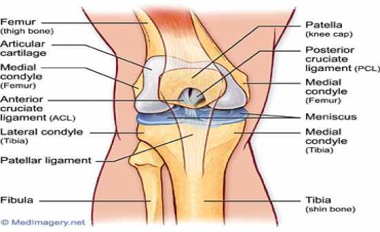
knee joint --> tibiofemoral joint
patellofemoral joint
lateral and medial menisci
transverse ligament
popiteal region
fibular (Lateral) collateral ligament
tibial (medial) collateral ligament
ACL
PCL

13 bursae: anterior
superficial infrapatellar
suprapatellar
prepatellar
deep infrapatellar

talocrural joint
posterior tibiofibular ligaments
anterior " "
medial (deltoid) ligament
lateral (collateral) ligament
calcaneal tendon (Achilles)